
Female Reproductive Anatomy: The Complete Guide
Photo by Deon Black on Unsplash
Our complete guide to male anatomy.
As you've probably noticed, we love chatting about sex and relationships at Green Condom Club! But first things first: knowledge of your anatomy (and your partner's one!) is essential, right?
Sure, most of us know the basics. But who can really tell how a clitoris works? What are the different functions of a vagina? And, do you know what happens precisely during each phase of a menstrual cycle?
And so, we thought that it was about time to write a complete guide to female reproductive anatomy.
What is the reproductive system?
The reproductive system consists of all the body parts that unable a woman to procreate and have sex. It comprises external genitals, which are the different parts of the vulva, and internal organs, the uterus, vagina, and uterine tubes. We'll also include breasts due to their essential role in breastfeeding (and so, reproduction!).
Structure of the female reproductive system
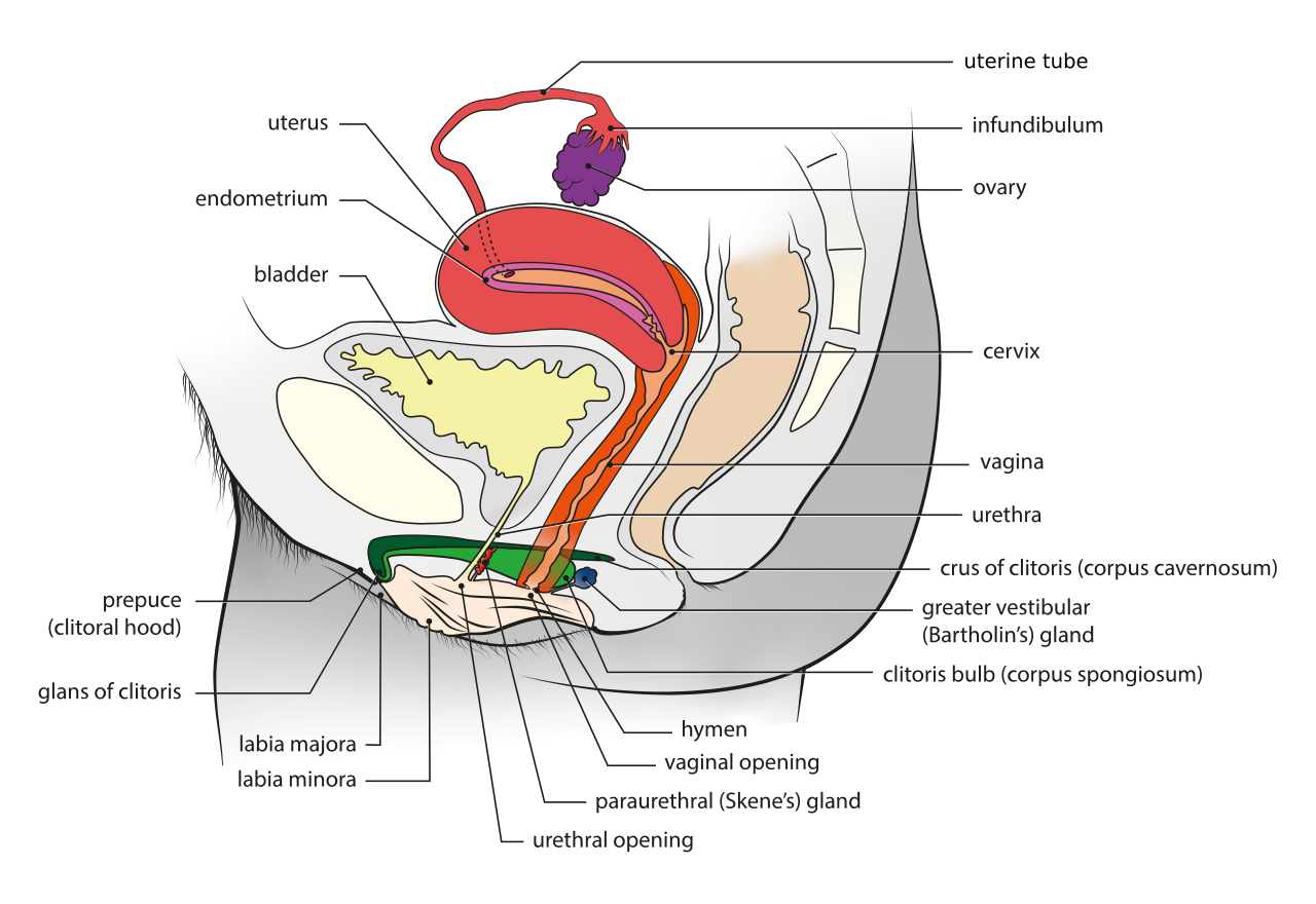
Female anatomy, perspective view. Anatomical charts of the genital system developped by Bioscope, HUG, DIP and Odile Fillod, published on the website Sciences, Sexes, Identités of Université of Geneva by R. Dewaele (Bioscope, Unige), J. Abdulcadir (HUG), C. Brockmann (Bioscope, Unige), O. Fillod, S. Valera-Kummer (DIP), www.unige.ch/ssi

Female anatomy, side view. Anatomical charts of the genital system developped by Bioscope, HUG, DIP and Odile Fillod, published on the website Sciences, Sexes, Identités of Université of Geneva by R. Dewaele (Bioscope, Unige), J. Abdulcadir (HUG), C. Brockmann (Bioscope, Unige), O. Fillod, S. Valera-Kummer (DIP), www.unige.ch/ssi
Female reproductive system parts and functions
The different parts of the vulva
Located in the perineum area, the vulva is composed of the external female genitals.

Female external anatomy, front view. Anatomical charts of the genital system developped by Bioscope, HUG, DIP and Odile Fillod, published on the website Sciences, Sexes, Identités of Université of Geneva by R. Dewaele (Bioscope, Unige), J. Abdulcadir (HUG), C. Brockmann (Bioscope, Unige), O. Fillod, S. Valera-Kummer (DIP), www.unige.ch/ssi
The mons pubis is the front part of the pubic symphysis (the joint between the two pubic bones), covered with hair.
The most external and visible part of the vulva is the labia majora, just below the pubis. It's a pair of folds of skin that covers the internal components of the female genitalia. In between the two labia majora, the labia minora (the inner folds of the vulva!) surrounds the entrance of the vagina.
In the continuation of the labia minora, there is the clitoral hood. Homologous with the male foreskin (and yes, girls have one too!), it's the upper part of the vulva covering the glans clitoris. A highly erogenous zone for women!
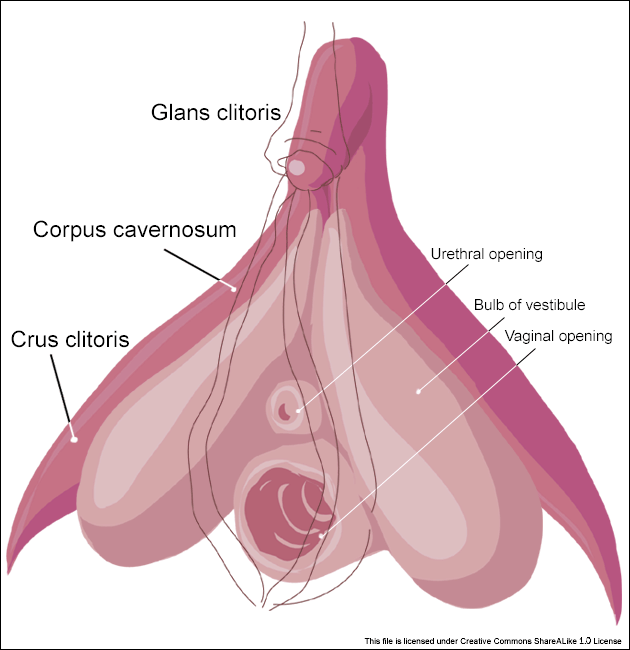
Internal anatomy of the clitoris by Amphis - Wikimedia Commons
What we imagine being the whole clitoris is actually the outer part, the glans, which is the size of a pea. Very few people know what this wishbone-like organ (also called bulboclitoral organ) really looks like: this 3D model will give you an idea.
The function of the clitoris has been a mystery for a long time, but we now understand that its sole purpose is female pleasure. To some extent, it can even be compared to the penis! In fact, the clitoris fills up with blood and swells during sexual excitation, which sometimes leads to an intense orgasm. That is, my friends, a clitoral erection!
Let's take a closer look at the different parts of the clitoris:
The two crura (legs) are attached to the pelvic bones. They are made of cavernous bodies (corpora cavernosa), which is an erectile tissue coated with a fibrous envelope called the albuginea. It allows them to swell during sexual arousal! More precisely, both legs are covered by the ischiocavernosus muscles: their contraction pushes the blood from the crura to the body of the clitoris. That creates a female erection!
The glans is the only part of the clitoris that is clearly visible. Depending on the woman, it measures from 2 mm to 1 cm. However, no less than 90% of this organ is actually internal! The glans constitutes its tip, containing more than 8000 nerve endings (the glans penis doesn't have more than 6000).
The body of the clitoris is formed by the union of the two crura, and is maintained by a ligament at the pubic symphysis.
Kobelt's plexus is shockingly often missing from anatomy books. Yet, it's an essential part! It consists of a network of veins that connect the bulbs to the body of the clitoris.
Under the crura, two bulbs surround the vaginal and urethral openings. Each bulb is formed of corpus spongiosum, an erectile tissue. Indeed, the bulbs play an important part in erection, too: the contraction of the bulbospongiosus muscles that covers them causes the clitoral erection. What is exactly happening? An erection occurs when the blood is pushed out of the bulbs towards the body part!
In between the bulbs is the vaginal opening, and just above it, the urethral opening, through which urine is evacuated from the urethra.
Bartholin's glands are two glands located on both sides of the vagina. From puberty to menopause, their role is to always keep the vaginal opening lubricated with a type of mucus.
The vulva vestibule is the part that comprises everything between the labia minora: it includes the urethral and vaginal openings, Skene's and Bartholin's glands.
The two Skene's glands are on either side of the urethra. They are often called the "female prostate" because their tissue is very similar to the male prostate. The role of these glands is to produce a liquid that has a comparable composition to the male's seminal fluid, but it comes out from orifices close to the urethral opening.
The different parts of the vagina
The vagina is a fibromuscular tube, starting from the vulva all the way up to the uterus. It has many functions:
Transporting the menstrual blood out of the body
Penetration during sexual intercourse (vagina/penis, masturbation, or any other combination)
Reproduction, as sperm travels through the vagina to reach an egg
Finally, childbirth. The newborn goes down through the cervix, the vagina, and the vulva to exit the body

Female internal anatomy. Anatomical charts of the genital system developped by Bioscope, HUG, DIP and Odile Fillod, published on the website Sciences, Sexes, Identités of Université of Geneva by R. Dewaele (Bioscope, Unige), J. Abdulcadir (HUG), C. Brockmann (Bioscope, Unige), O. Fillod, S. Valera-Kummer (DIP), www.unige.ch/ssi
The hymen is a membrane located at the entrance of the vagina. Contrary to what we imagine, it doesn't entirely cover the opening as it needs to let the menstrual blood pass through. Symbol of virginity, the hymen doesn't have any physiological function, and only half of women actually bleed during their first sexual intercourse.
The different parts of the uterus
The cervix is a cylindrical organ that separates the vagina and uterus. It has glands, called the cervical glands, that produce cervical mucus. Actually, its texture tells a lot about the stage of a woman's fertility: around the time of ovulation, the mucus is often compared to egg white as it turns abundant, fluid, transparent, and runny. That's what allows spermatozoids to travel smoothly towards the uterine tubes!
However, when a woman isn't fertile, cervical mucus thickens to prevent germs from entering the vagina. Observing these kinds of signs the body gives is a great way to control fertility, whether you want to get pregnant or avoid it! That's what the natural contraception method, called the rhythm method, is all about.
During pregnancy, the cervix is "blocked" by antibacterial mucus, evacuated when labor starts. Contractions of the uterus allow dilation of the cervix, thus the passage of the baby!
Above the cervix is the uterus, a sort of pocket leading to the uterine tubes and the ovaries. During pregnancy, the uterus hosts the fertilized egg - that is, the embryo - until its complete development. It goes from 8 cm to 35 cm in length, because it gradually stretches as the baby grows! Two months after birth, muscles should get back to their usual size.
But there is so much more to know about the uterus! A mucous membrane covers the uterine lining: it's the endometrium. Its thickness varies according to the menstrual cycle. Indeed, at the beginning of the cycle (first day of period = first day of cycle), the endometrium progressively thickens to allow a potential embryo to be implemented. If an egg isn't fertilized, the endometrium is eliminated during menstruation; but the endometrium contributes to making the placenta if it gets fertilized.
Starting from the uterus, the two uterine tubes (12 to 15 cm long!) end with the ovaries. Their function is essentially linked to procreation: the spermatozoids travel all along the tubes to reach the pavilion, where an oocyte is waiting to be fertilized.
The pavilion is the end of the uterine tube. Every month, an oocyte is expelled by the ovaries, and remains in the pavilion until it gets fertilized. If so, the egg will move towards the uterine cavity to develop.
The ovaries are the female reproductive organs, comparable to the male testicles. They produce sex hormones: progesterone and estrogen, and are responsible for developing secondary sexual attributes such as the voice, breast, body shapes, and hair.
The most inferior part of the pelvic area is the perineum, a muscular zone between the coccyx, the sacrum, and the pubis. Its function is to support the organs located in the small pelvis: the rectum, the uterus, and the bladder, thus controlling urinary and fecal continence.
Then, you can imagine how essential the perineum is for pregnancy and childbirth: it supports the pelvis and the uterus while allowing them to enlarge progressively. It loosens up after childbirth, which is why it's fundamental to reeducate the pelvic floor muscles to avoid prolapse and incontinence later on in life.
The urinary system
The urethra duct carries urine from the bladder out of the body, through the urethral opening.
The bladder is the pocket-like organ that stocks up urine, previously produced by the kidneys. Ever wondered how we can hold it? Thanks to the bladder sphincters!
The end of the digestive system
The last part of the digestive system is the rectum, which marks the end of the colon and leads to the anus. In the shape of a curve, it measures no less than 15 cm! The rectum comprises two parts: the rectal ampulla, which stocks the fecal matter before exiting the anus, and the anal canal, which allows continence.
The anus is the very end of the digestive tract! This orifice is located just after the rectum and regulates defecation, thanks to the sphincters, which play an essential role in controlling continence.
The breasts
Let's finish with our beautiful paired organs: breasts! Externally, they are composed of a nipple and an areola (the pigmented skin that surrounds the teat), and internally, of fatty tissue and mammary glands, which produce milk to feed newborns.
How does the reproductive system function?
The different phases of a menstrual cycle
The follicular phase (day 1 to 14): day 1 corresponds to the first day of menstruation. After bleeding for several days, the endometrium is reformed to prepare for a potential pregnancy.
Ovulation (day 14): on a cyclical basis, women produce gametes called oocytes, namely eggs that have not reached maturity yet. It means that from puberty to menopause, every 28 days (approximately), an ovary releases an oocyte during ovulation. If a spermatozoid does not fertilize it, it survives only a couple of days.
The luteal phase (day 14 to 28): if there isn't pregnancy, the uterus prepares to evacuate its internal lining, called the endometrium. That's what provokes periods! Thus a new cycle begins…
Everything you always wanted to know about… ♥
What is squirting?
Have a look at our article on the topic!
What is the function of the clitoris?
The history of the clitoris is a fascinating one, and it says a lot about the perception of female sexuality! To know more about the only organ that is meant for pleasure, I advise you to watch Maria Rosok's TEDx Talk, which will tell you everything about "the unknown greatness of the clitoris".
What is menopause?
Menopause is a natural event that happens to women around 50 years old. From that time, they stop being fertile, having their periods, producing eggs and reproductive hormones (estrogen and progesterone) altogether.
THE LATEST BLOG ARTICLES


Let's be honest: what is the first thought that comes to your mind when you hear about scheduling sex? Boring. Laborious. So-not-sexy. Yet, many sexologists swear by this practice to cultivate intimacy within a relationship.